Our lens probes, custom designed for in vivo imaging with nVista, enable you to visualize Ca2+ activity in hundreds of neurons in the hippocampus in freely behaving rodents. Study place cell coding, spatial navigation, learning and memory and more!
https://www.youtube.com/watch?v=4CmP4QHrpCQ
https://www.youtube.com/watch?v=LORIS9BVsjM
The nVista system has opened a whole new avenue of research for us. We found it relatively easy to adopt for recording in the hippocampus of behaving mice, and are amazed at the wealth of data it provides and consequently the power this approach adds to our studies on hippocampal function.
Description
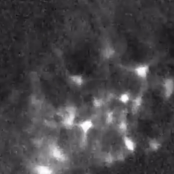
Animal Model: Mouse Brain Area: Hippocampus, CA1 Indicator: GCaMP3 Notes: Decoding CA1 place fields in a linear track. Courtesy Of: Ziv, et. al., Nature Neuroscience, 2013.
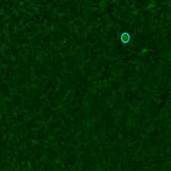
Animal Model: Mouse Brain Area: Dentate Gyrus Indicator: GCaMP6 Courtesy Of: Stuber Lab, UNC